Before using the Xepr program the spectrometer needs to be turned on. The order is as follows: chiller, main power supply, main spectrometer unit, and computer. Details in Manual EPRI file.
PDF FILE | MANUAL |
| MOVIE 1 | 1. Connecting to spectrometer, creating experiment and tuning Connecting to spectrometer. Computer needs to be connected with the control unit of the spectrometer. It’s done once after turning on the Xepr program. Creating basic type experiment with field sweep only (without gradients). Before starting to gather data spectrometer needs to be informed what type of experiment will be made. The spectrometer switches on or of the parts which will be used or not. Tuning is necessary that all applied energy from microwave source will get to the resonator |
| MOVIE 2 | 2. Basic experiment - field sweep Basic EPR experiment with all possible settings is showed. There is no possibility of turning on gradients or performing other 2D experiments not imaging experiments Multiple field scans show the influence of increasing of the modulation amplitude beyond allowed limits which results in line broadening |
| MOVIE 3 | 3. Experiment with gradients Experiment with gradient unit turned on is prepared. The influence of rising gradient strength is shown. |
| MOVIE 3a | 3a. Experiment with gradients and gradient calibration Additionally, to the previous experiment an automatic gradient calibration procedure is shown. Should be used only when applied gradients shift the center of the EPR signal. |
| MOVIE 4 | 4. 2D imaging and reconstruction to slices Imaging type experiment is created. 2D spatial imaging experiment is performed. The recorded projections undergo deconvolution and filtered back projection resulting in the final 2D image. |
| MOVIE 5 | 5. 3D imaging and reconstruction to slices 3D experiment is performed with similar reconstruction path as it was for 2D image. Obtained slices should be stacked one above another to get the 3D image. |
| MOVIE 6 | 6. Example 1 - 3D reconstruction from slices Bruker calibration phantom 6 dots of DPPH in a cube, separated by 5 mm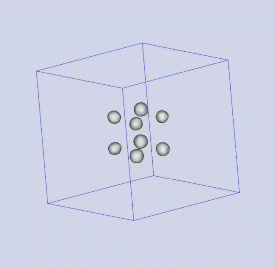 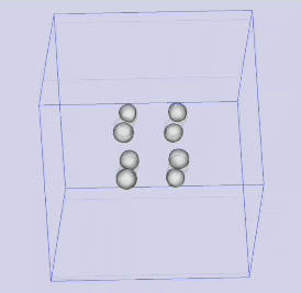 |
| MOVIE 7 | 7. Example 2 - 3D reconstruction from slices – Polydopamine covered reduced graphene oxide aerogel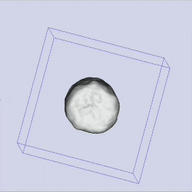 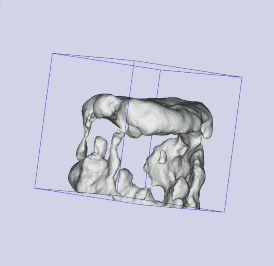 RGO Aerogel: 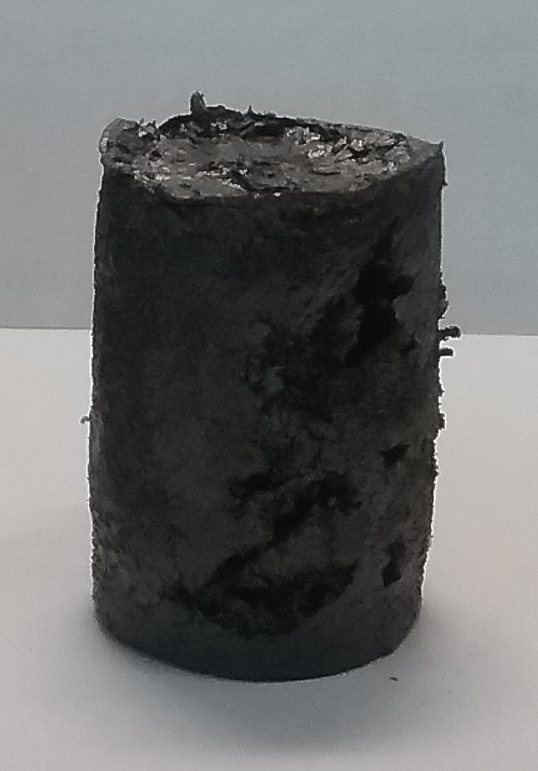 |
RGO Aerogel
EN