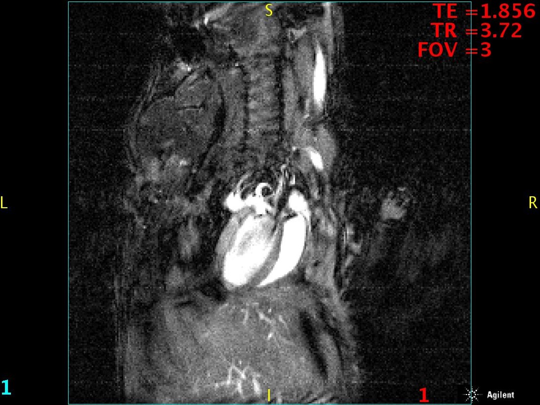
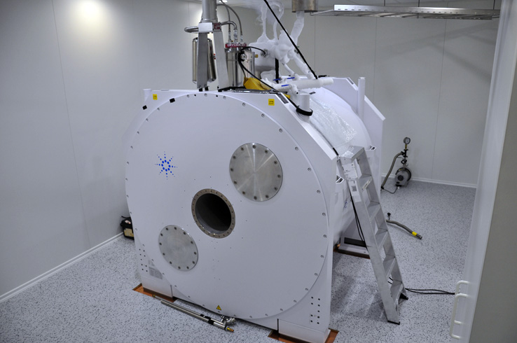
Scanner MRI 9.4 T 310 mm
Wide bore 310 mm 9,4 T superconductive magnet.
System has 2 gradient system:
- Coil 210 mm bore with gradient 1 m|T/m/A
- Coil 120 mm bore with gradient 2 mT/m/A
Excitation-receiver coils:
- Birdcage outer dimension 210 mm – for large animals (rabbits), rats
- Birdcage outer dimension 120 mm – for rats, mice
- Millipead coli 30 mm diameter – to biological samples, millipead allow get images with better resolution then birdcage coil
- Millipead coil 40 mm diameter – to biological samples, millipead allow get images with better resolution then birdcage coil
- Surface coil – to investigate animal brain
MRI scanner has full physiological monitoring with animals holders.
System supports a full range of MRI pulse sequence including echo planar imaging and diffusion tensor imaging.

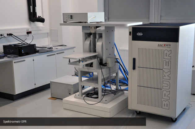
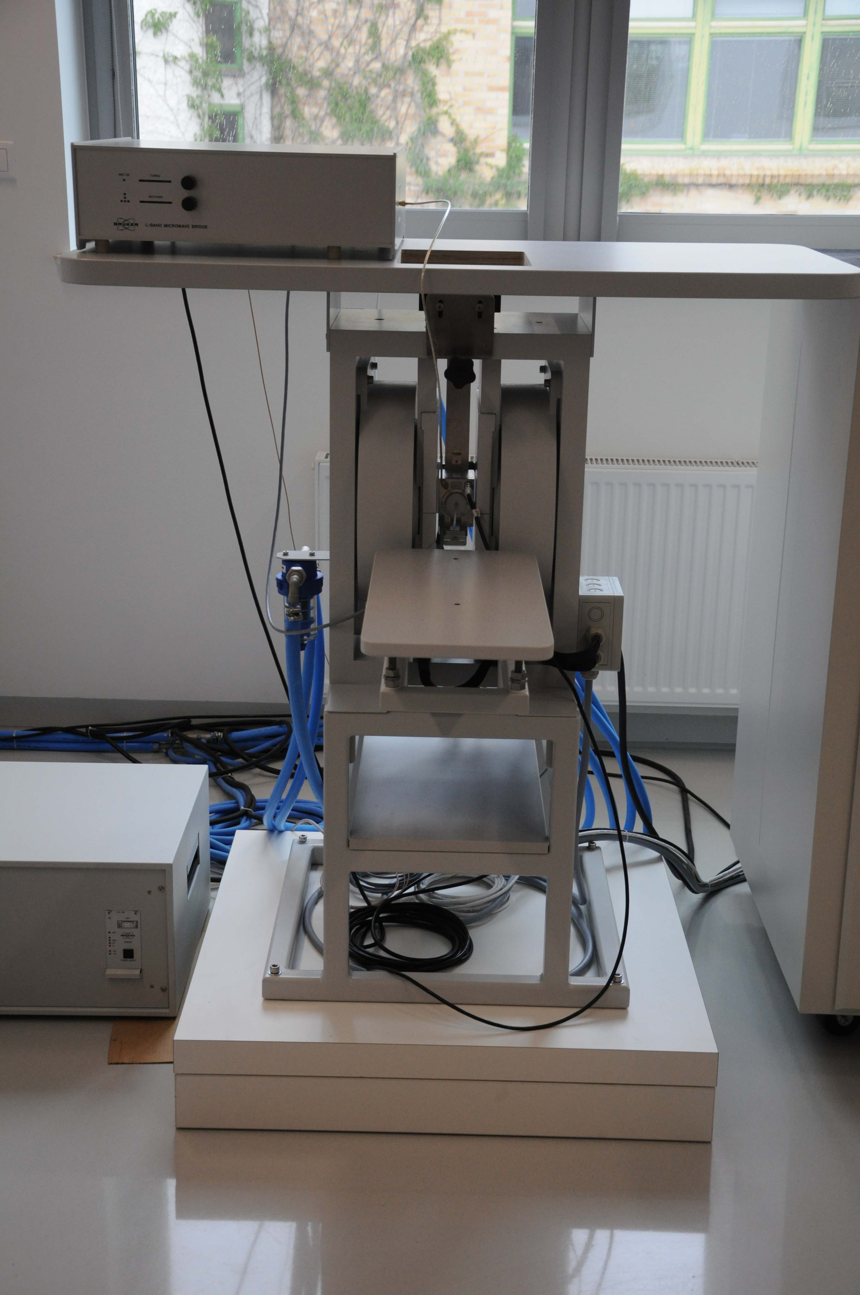
Electron Paramagnetic Resonance (EPR)
Is a spectroscopic technique based on the absorption of the electromagnetic radiation in the range MHz – THz through the electrons with unpaired spins, when sample is placed in the external magnetic field and resonance conditions are fulfilled. In the EPR Imaging (EPRI) additionally the sample is placed in an external magnetic field gradient which allows the spatial information to be obtained. For image reconstruction this technique uses similar algorithms as Computer Tomography. NanoBioMedical Center owns the Bruker ELEXSYS-II E540, continuous wave, 1GHz spectrometer, equipped with E540R23 resonator (23 mm access tube) which allows in-vivo imaging of small animals e.g. mice.
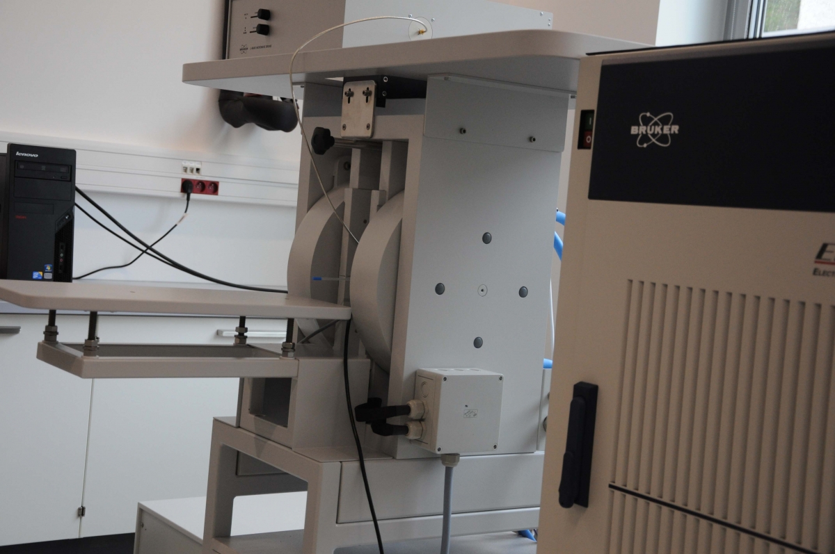
Bruker ELEXSYS-II E540 spectrometer
MANUAL WITH EXAMPLES
Bruker ELEXSYS-II E540 spectrometer
MANUAL WITH EXAMPLES

Equipment:
- Bruker ELEXSYS-II E540 (CW, 1GHz) spectrometer equipped with E540R23 resonator
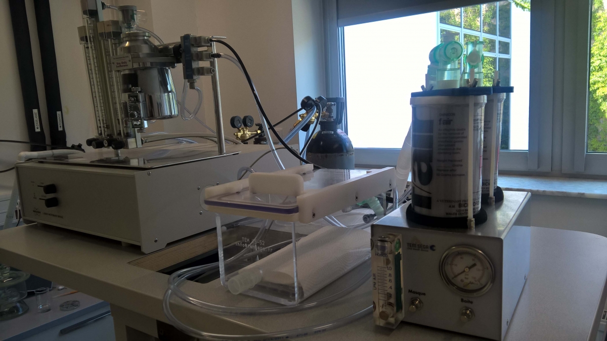
- Mouse anesthesia system
- Animal handling system for EPR measurments
- Optical oximeter Fibox4 (PreSens), with PST7 probe
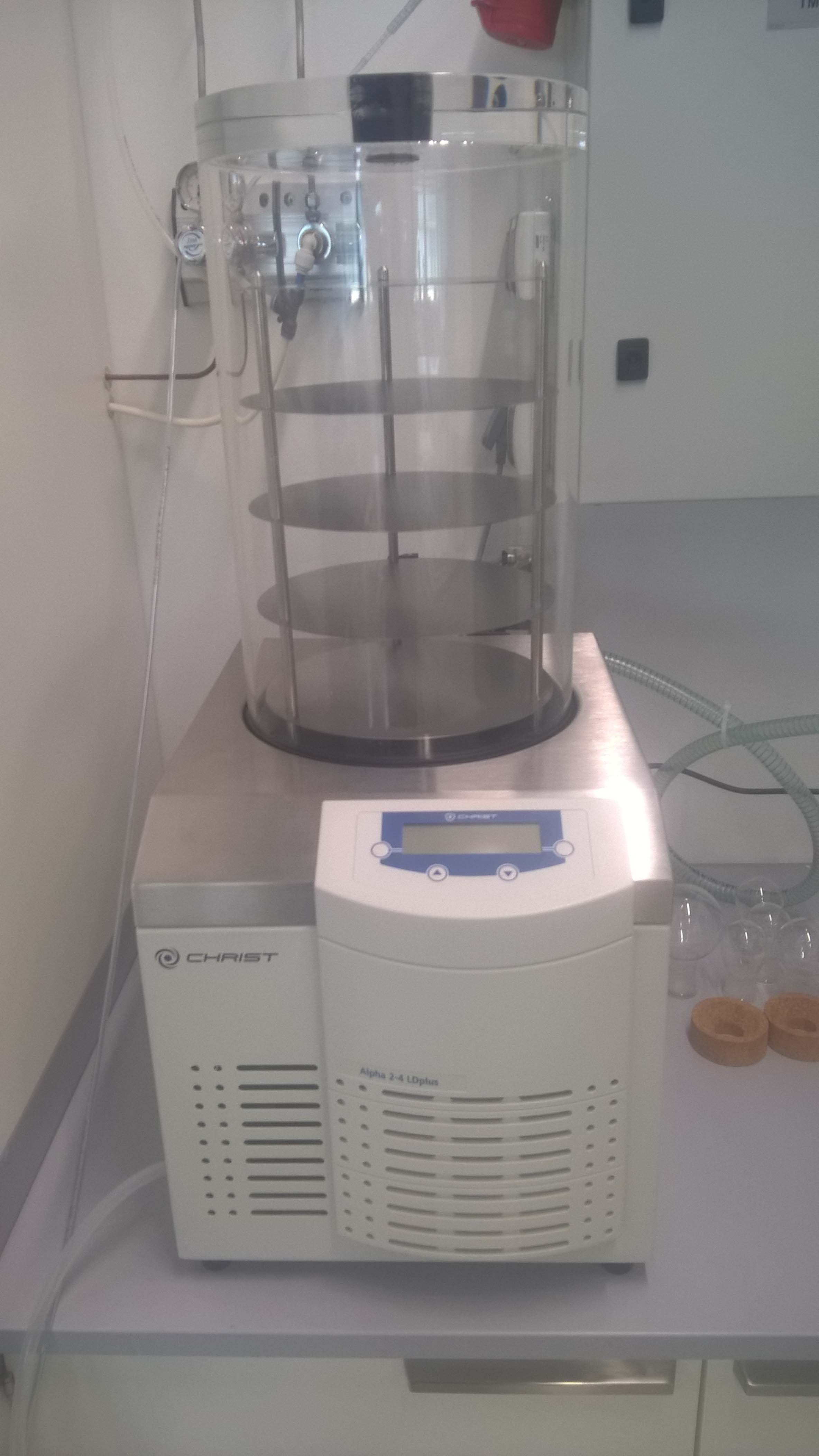
- Lyophilization Alpha 2-4 LDplus
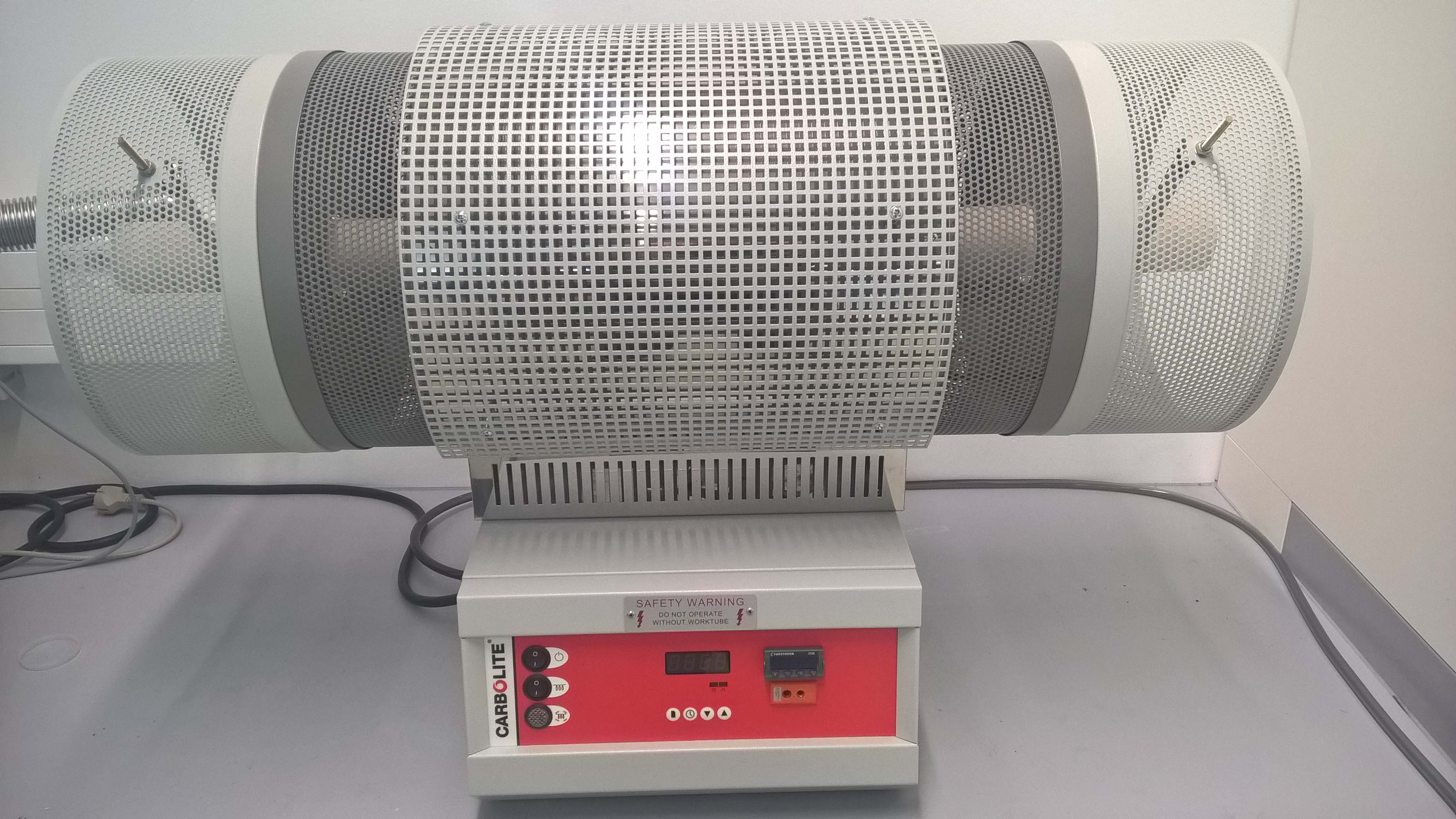
- Furnace Carbolite (1600C)
- PreSens Fibox 4 fiber optic trace oxygen transmitters - oximeter
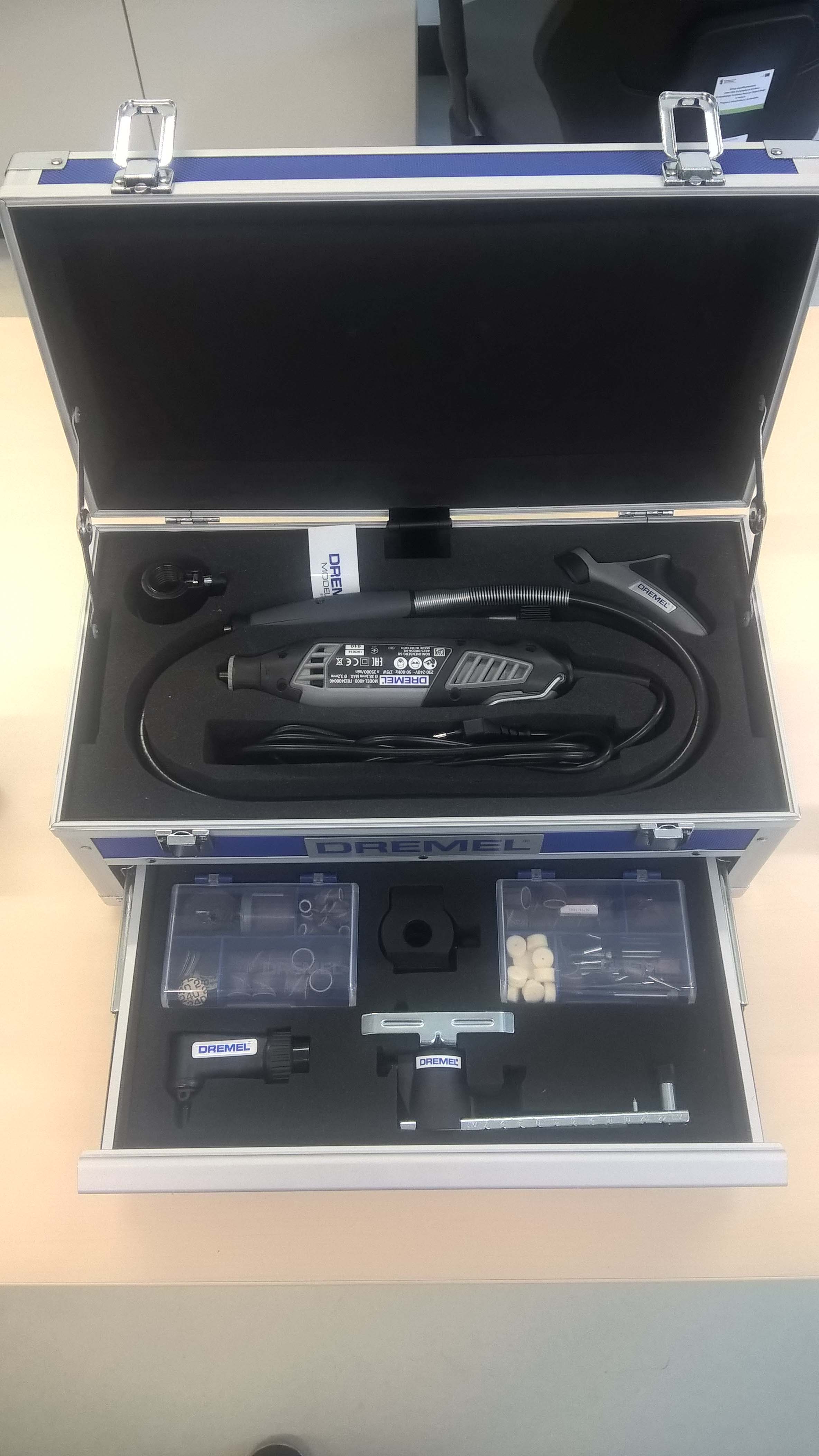
- Dremel Platinum Edition 4000-6/128 Corded Multitool (175 W), 6 Attachments, 128 Accessories
Advantages of EPR technique:
- Enables the detection of paramagnetic centres: persistent radicals in biological materials, drugs, tissues, blood, and ions,
- Enables the measurements of oxygen concentration in liquids especially blood by the use of dedicated radical spin probe,
- Enables the study of spin dynamic in biological systems,
- Is useful in the studies of ionizing radiation on biological samples.
EPR imaging technique allows:
- Spatial distribution of radicals in the sample (2D and 3D spatial imaging). In spectral-spatial imaging 2D, 3D, and 4D imaging is possible,
- Monitoring mobility, polarity, molecular oxygen levels (pO2) and pH,
- Monitoring of drug release from nanostructures, polymers etc.,
- Study of diffusion of radicals in porous materials
Example applications of EPR imaging:
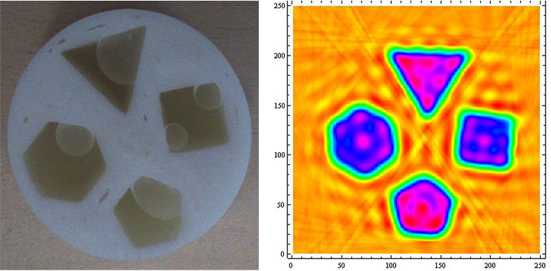
PLA phantom of different shapes with 2.5mM TEMPO solution in water and reconstructed 2D image
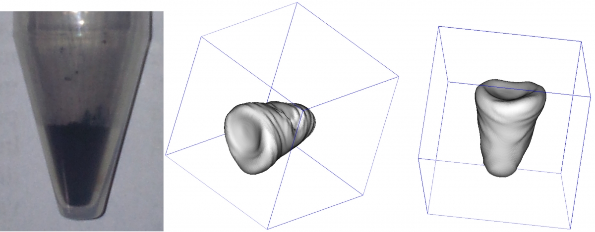
Polidopamine powder in Nunc™ 15mL conical sterile polypropylene centrifuge tube and 3D (spatial) image reconstruction