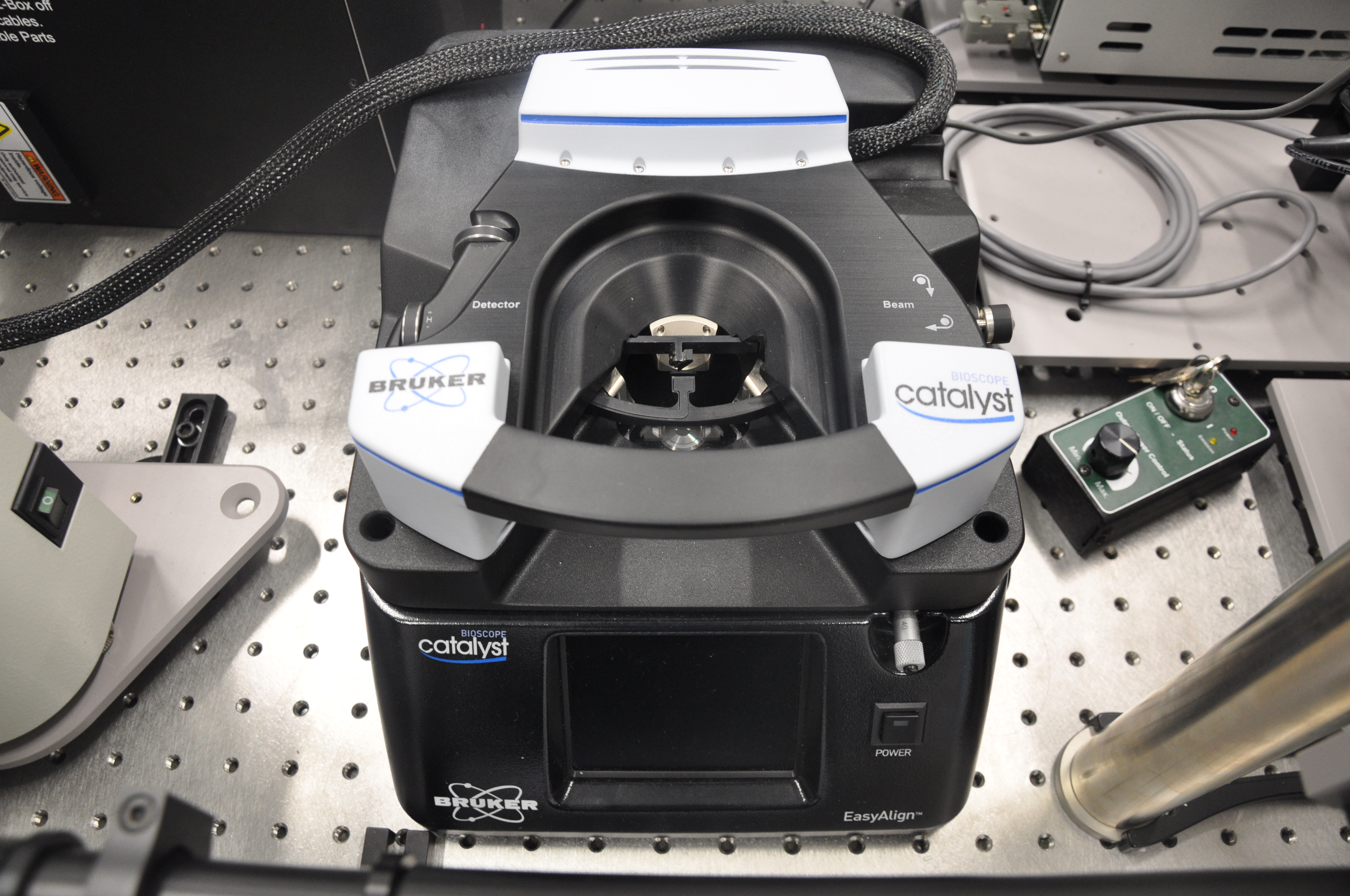
The microraman spectrometer and the bioscope Catalyst
The microraman spectrometer combined with the bioscope allow simultaneous measurements of molecular dynamics (including surface mapping) and surface physical properties using a multimode atomic force microscope (incorporating latest PeakForce Tapping innovation).
Important parameters:
Temperature range -195 0C till +600 0C
Laser wave length:785 nm,633 nm, 514 nm
- Spectral resolution better than 1 cm-1
- Spectrometer mode: surface mapping (streamline imaging),
- Depth mapping
- AFM (multimode system)
- Life cell imaging
Application: biology, material science.
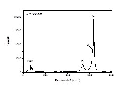
Raman spectra collected from the multiwall carbon nanotube shows three characteristic bands: G, D and RBM. Band G is related to bulk carbon structure, band D to disorder in the carbon nanotube and band RBM to a radial breathing mode. A wave number of the RBM depends on diameter of the tube.
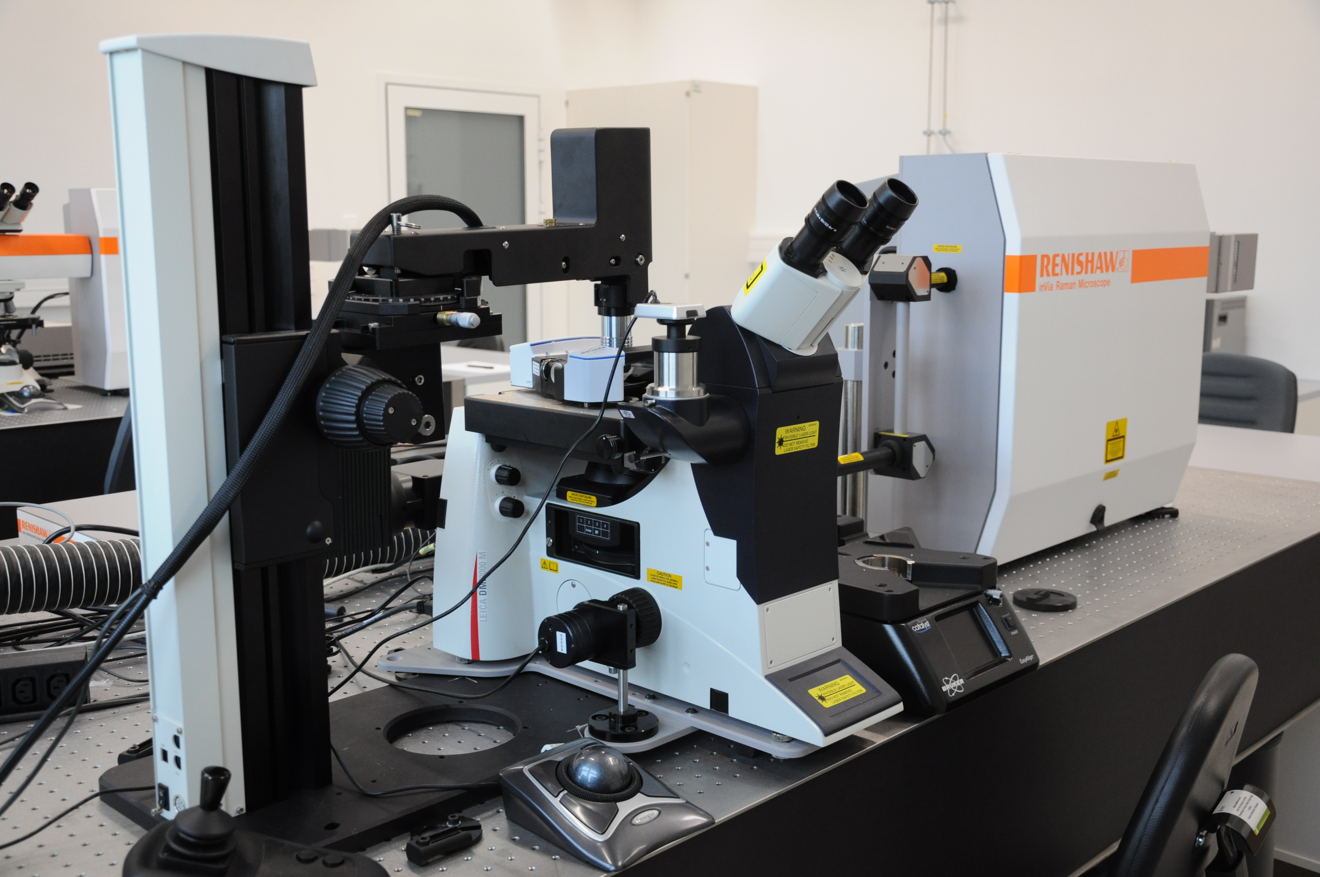

A microraman spectrometer and a NT-MDT SNOM microscope
Important parameters:
- Laser wave length:633 nm,514 nm, 488 nm
- Spectral resolution better than 1 cm-1
- Spectrometer mode: surface mapping (point imaging),
- Depth mapping
Modes:
- Share force
- Transmission
- Reflection
- AFM (multimode system)
- Thermal conductivity
Application: material science, biology

Presented in Fig.2 result of the surface mapping mode shows a nonuniform graphene sample.
The 2D peak related to number of layers of a graphene flake, mapped over a sample surface, reflects change in the sample’s thickness.
Optical microscopy
Microscopes and equipment
Zeiss:
- scanning fluorescence microscope LSM 780 NLO,
- halogen + fluorescence lamp + filters
- CW lasers: 405, 458, 488, 514, 561, 633 nm
- Two photon excitation (Chameleon 680-1080nm, 140 fs),
- spectral module,
- fluorescence correlation spectroscopy (FCS – ConfoCor 3)
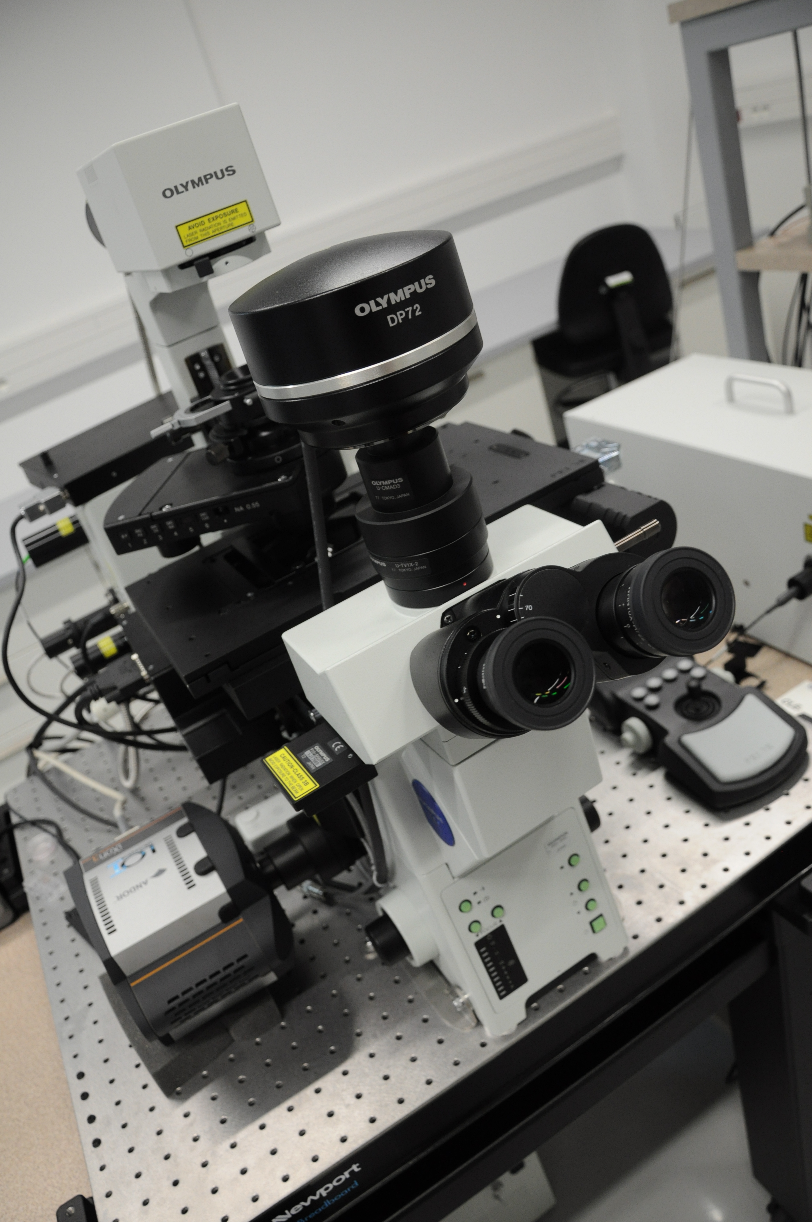
Olympus:
- scanning fluorescence microscope FV 1000,
- halogen + fluorescence lamp + filters
- CW lasers: 405, 457, 473, 488, 514, 561, 638 nm
- spectral module,
- FLIM (485, 635 nm),
- TIRF (Andor camera),
- FCS (Picoquant)
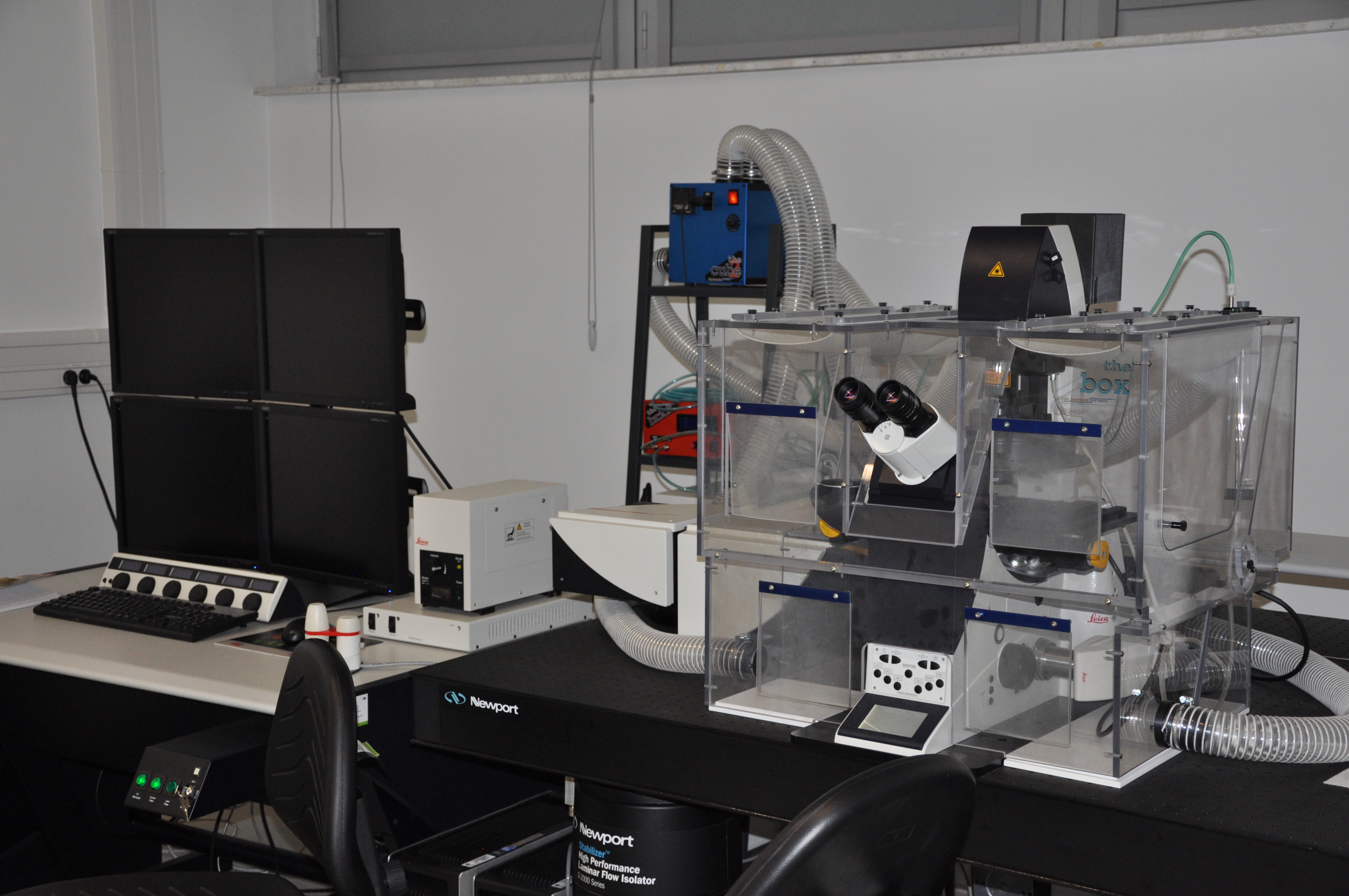
Leica:
- scanning fluorescence microscope
- halogen + fluorescence lamp + filters
- STED (super-resolution),
- white laser 470 – 670 nm,
- CW lasers: Ar, 458, 476, 488, 496, 514 nm
- spectral module,
- incubation chamber (temperature, CO2)
- FCS (Picoquant)
The optical microscopy lab provides instruments for studying the structure, dynamics and optical properties of matter in the nano- and micro-scale.
- Each of the three microscopes may serve as a regular optical microscope operating with magnification of ~1000x both in the bright field and fluorescence mode.
- Each of the three microscopes may work in the mode of the laser scanning fluorescence microscope in order to obtain precise images of fluorescently labelled samples. A broad spectrum of excitation wavelenghts is available.
- Each of the three microscopes may perform spectral measurements which allows measurements of fluorescence spectrum of single molecules or of sub-micron regions of the sample.
- Each of the three microscopes can measure the kinetics of fluorescence fluctuations (fluorescence correlation spectroscopy – FCS) which allows for measurements of diffusion coefficients of fluorescently labelled particles in extremely low concentrations (nanomolar).
- Olympus microscope has two special options: FLIM (fluorescence life-time imaging) and TIRF (total internal reflection imaging).
- Zeiss microscope is equipped with a tunable femtosecond infrared laser used for two-photon excitation.
- Leica microscope is equipped with a tunable visible laser (“white laser”) and the STED super-resolution option (stimulated emission depletion) extending the resolution to ~40 nm range.